Newsletter 07-2016
13th July 2016

Introducing our new Chief Executive Officer
In June this year I started as CEO of Scannexus. As probably many of you already know I am also Dean of the Faculty of Health, Medicine and Life Sciences and Vice Chair of Maastricht University Medical Centre.
I have got many reactions on this new parttime job (officially 0.8 Dean and 0.2 CEO). Some colleagues immediately understood it and some others asked me if the job as Dean was not busy enough. Yes, the job as Dean is busy enough and the fact that I am CEO means that the other members of the board and the supportive staff at FHML have more work.
So, why I accepted then the job? I see Scannexus as a great platform with many opportunities. My opinion is that we all together on the Brightlands Maastricht Health Campus should work together to make our Campus stronger and even more exciting than it already is. I also see that there can be frictions between business and science on research platforms like Scannexus. Since several years we are talking at the MUMC about sharing infrastructure and we are working at research platforms, because we see that as the only future for expensive infrastructure. Sharing is in our vision the only way to keep expensive infrastructure up to date. So, when the possibility came of becoming CEO, my decision was quite easy. I saw a nice challenge that was aligned with the MUMC vision and mission and I have the opportunity to stimulate collaborations on our campus and in the region.
Albert Scherpbier
CEO, Scannexus
Brains Unlimited Pioneer Fund 2016
In April of this year a 4th call of the Brains Unlimited Pioneer Fund was opened. After a great interested and many applications, the Advisory Board has approved 9 new exciting projects! We would like to congratulate the scientist with the allocation of the Brains Unlimited Pioneer Fund.
In the previous three rounds 24 were applications that were approved, many projects are currently working hard on their studies or already finalizing their projects.
The Brains Unlimited Pioneer Fund supports research, carried out by UM researchers in the Scannexus MRI facility. The Pioneer Fund makes money available for innovative pilot studies which have a collaborative nature. These studies aim to generate a proof-of-evidence that substantiates an idea and increases the likelihood of further grant funding for follow-up research.
For more information on the Brains Unlimited Pioneer Fund, please visit the website of the University Fund Limburg or 'click here'.
The Brains Unlimited Pioneer Fund has been made possible by the University Fund Limburg/SWOL. If you would like to support the good cause that is served by this specific fund, please contact Jos Kievits, director University Fund Limburg for the possibilities (jos.kievits@maastrichtuniversity.nl).
'Click here' to see the approved Brains Unlimited Pioneer Fund applications.
Insight into the functional brain networks in participants with type 2 diabetes and pre-diabetes by Frank van Bussel, PhD
Participants with type 2 diabetes have altered functional brain networks. This alteration is already apparent in the pre-diabetic stage, hinting at functional reorganization of the cerebral networks as compensatory mechanism for cognitive decrements.
Four years ago, van Bussel started his PhD in cooperation with The Maastricht Study to gain more insight into the neuronal mechanism underlying cognitive decrements in type 2 diabetes. One part of his research focused on the functional MRI (fMRI) data of The Maastricht Study, which was acquired by Scannexus on a 3 Tesla MRI scanner. Besides participants with type 2 diabetes and healthy participants, we selected also matched participants with pre-diabetes from the MRI data to investigate whether we were able to observe functional network alterations (if present) in an early diabetic stage. Van Bussel: “Thanks to Scannexus that they have scanned many participants in such a short time frame for The Maastricht Study, which makes it easier for me to select the participants from a large MRI study pool and achieve my results before the end of my PhD.”
Importance
Diabetes is not only connected to cardiovascular disease, but also affect the brain in terms of cognitive decrements, accelerated cognitive decline and a twofold increased risk to develop dementia, including Alzheimer’s disease. Unfortunately, the consequences of these complications could lead to a decrease of the quality of life, including, in a later stadium, less self care and less abilities to maintain social contacts. Therefore, it is important to gain more insight into the neuronal mechanisms underlying cognitive decrements in diabetes, which hopefully leads to the development and evaluation of better treatment or intervention strategies to delay or even prevent the worsening cognitive decrements in diabetes.
The role of MRI
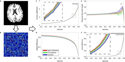
MRI could provide us more insight into the underlying mechanism and, more important, it could be noninvasive for the patient. These are the reasons that we used the resting state fMRI data, acquired by Scannexus, to investigate whether we were able to identify alterations in the functional brain networks. A total of 47 participants with type 2 diabetes, 47 pre-diabetes, and 47 control participants were involved in this study. To achieve the final result, a number of (pre-)processing steps are needed (Figure). In brief, starting with the brain (Figure A), we extract a brain graph (Figure B, each row and column represents a brain area) from the fMRI data. The brain graph represents which brain areas are functional connected to each other (red represents high correlation between two brain regions, which are thus functional connected; blue represents low correlation between two brain areas, which are thus less or not functional connected). Finally, brain network measures (Figure C-F) were calculated from the brain graphs and characterize how well the brain is organized. This study reveals altered functional brain networks in participants with type 2 diabetes as well as pre-diabetic participants, indicative of a more efficient cerebral organization, hinting at compensation for cognitive decrements in terms of functional reorganization of the cerebral networks.
Next step
The outcomes of the presented study provide new insights and have the potential to serve as an early MRI biomarker for subtle cerebral alterations in relation to cognitive decrements. Nevertheless, future studies are needed to confirm the potential of this MRI biomarker and for this it is important that the participants undergo one or more of the same fMRI examinations. The idea of The Maastricht Study is to plan a follow-up round and Scannexus has already demonstrated that they are able to cope with the MRI examinations of a large group like ‘The Maastricht Study’ participants. Furthermore, Scannexus has the facilities and expertise that leads to a successful cooperation.
“Scannexus has successfully demonstrated to cope with many MRI examinations and MRI data of large cohort studies”
European IDEA User Group Meeting 2016
Maastricht is proud to be hosting this year’s European IDEA User Group Meeting, at Oxfordlaan 55 on 20-21 September 2016. This biannual event is held at a different site each time, and intended for those of the MR imaging community with experience and/or interest in IDEA programming for the Siemens MRI scanner platforms.
The workshop will be organized jointly by Siemens and the Faculty of Psychology and Neuroscience, with support from Scannexus.
Scientists from Siemens Healthineers will be in attendance and will discuss new and planned features of IDEA. Similar to previous users meetings, participants will determine a majority of the program content based upon their work chosen to present.
The purpose of the IDEA User Group Meeting is to:
- Provide a forum for representatives from various user groups to present and discuss challenges encountered while utilizing the IDEA development environment along with unique and innovative solutions;
- Provide information about new and planned features of IDEA along with hands-on sessions;
- Exchange experiences and get to know other IDEA users and therewith facilitate the collaboration of user groups.
Scannexus will be sponsoring the event with a EUR 2000 contribution to the dinner night, and provide scanner access for live demonstrations.
Online registration will open in late June / early July. Links to the registration page will posted on the website of FPN-BIC, Scannexus.nl and the Siemens IDEA online portal.
30 years of MRI research by Christopher Wiggins, PhD
Recently I was reminded of just how long I have been working in the field of MRI. The reminder came about in an article in the Scottish television company STV’s news edition entitled “First MRI goes on display in Aberdeen hospital art gallery”. The article described how this system, the Aberdeen Mark One, had just been reassembled in the Suttie Arts Space Gallery. The Mark One - one of the first three whole-body MRI systems ever constructed – is notable in MRI history in several facets. Particularly it was the scanner where the first ever diagnostic images were made, as well as being where the basis of most modern MRI techniques – ‘Spin-Warp’ phase encoding – was invented and developed.
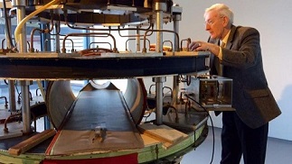
First MRI: Creator Eddie Stevenson with his machine. STV
My start in the field was somewhat accidental. I was an undergraduate student at Edinburgh University, and was trying to decide which career path to take. With both a father and a maternal grandfather having been research physicists, research always seemed the most natural path. However I decided that it would be good to get some experience in a research lab before my final year, so that I could get the feel of how it felt in practice. I had once toured the Medical Physics department at the Royal Infirmary in Edinburgh (where there was an early commercial MRI system, an M&D 800, which was from a spin-off company from Aberdeen) and liked the idea of applying physics to medicine. I therefore wrote to the Medical Physics department in Aberdeen, asking whether they had a position for a student for the summer. They wrote back inviting me for an interview, as they had a position for a student in the cyclotron unit connected to their PET (Positron Emission Tomography) scanning centre. I duly took the train to Aberdeen while reading up on cyclotrons (I believe it was the book The Cyclotron by W.B. Mann, published by Methuen. As I recall this was pretty much a do-it-yourself manual for how to construct one!). However when I got to the department all they talked about was MRI. Later I found out that there had been a severe fire in the cyclotron unit, and so any work for a student was gone (other than sweeping up the ashes, I guess) and so they decided that MRI was a better option. Thus I spent the summer working under Dr. Tom Redpath, measuring gradient non-linearities, developing a pneumatic belt for respiratory measurements, and using the latter to try to implement a technique called ROPE. One of the PhD students at the time is now well known in the MRI community, both in general and particularly in the Netherlands: one David Norris (now at the Donders in Nijmegen). After completing my degree in Edinburgh, I then attended the Masters in Bio-Medical Physics in Aberdeen, completing the research part of my thesis – Techniques for Fat-Water Discrimination - under David Lurie. I then worked there for a year as a Research Assistant in the Bio-Medical Physics department.
So what was this system like? 'Click here' to read the complete article.
Annual meeting of the ISMRM in Singapore
In May Scannexus joined the 24th annual meeting of the ISMRM in Singapore. Singapore is a beautiful city and we enjoyed our time. After the opening reception we had an informal Scannexus get-together with researchers from Maastricht surroundings. It was a great opportunity to get to know each other and to exchange thoughts and ideas while enjoying some food and drinks.
Next morning we started to explore the congress and to look for exciting developments. The Siemens 7 Tesla clinical machine, Terra, will enter the market very soon, so we were also keeping an eye out on future clinical applications at ultra- high- field.
We would like to highlight some work of researchers who collected their data on our MRI scanners and/ or are welcome guests to our facilities. Tamar van Veenendaal proudly presented her method to reveal spatial neurotransmitter networks using 7 Tesla spectroscopic imaging data. Another 7 Tesla user, Sriranga Kashyap, presented an alternative approach for collecting anatomical data to avoid the geometric dissimilarities- resulting from the distortions at ultra-high field -between (cortical-depth dependent) fMRI and anatomical data.
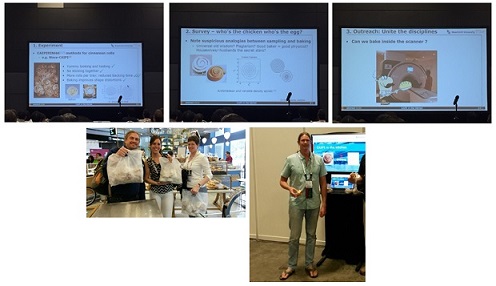
Recent work on the 9.4 Tesla was presented by Francisco Lagos. A specialized coil was used to achieve 450μm resolution diffusion imaging of the whole human post mortem brain. Desmond Tse presented his optimized workflow for high resolution GRE imaging at the 9.4 Tesla using SMS-pTX spokes excitations. We are grateful to him and Benedikt Poser for all the work they did to uncomplicated scanning at the 9.4 Tesla. At the ISMRM power pitch theatre Benedikt disclosed that MRI can be as (un)complicated as baking and that the use of CAIPIRINHA principles can result in lovely cinnamon rolls. To support this insight we brought 50 cinnamon rolls for the audience as a treat.
Ultrahigh Field Magnetic Resonance: Cutting Edge Technologies Meet Clinical Applications
The development of ultrahigh field magnetic resonance (UHF-MR) is moving forward at an amazing speed that is breaking through technical barriers almost as fast as they appear. UHF-MR has become an engine for innovation in experimental and clinical research. With almost 40,000 MR examinations already performed at 7Tesla, the reasons for moving UHF-MR into clinical applications are more compelling than ever.
That makes this a perfect moment for a topical Special Issue on "Ultrahigh Field MR: Cutting Edge Technologies Meet Clinical Applications", just published (June 2016) in the journal of Magnetic Resonance Materials in Physics, Biology and Medicine (MAGMA), a catalyst of novel imaging methodology and its (bio)medical applications, which ranks among the top 30 journals in the field of imaging/radiology.
This special issue may be of interest to you. Scannexus is mentioned in three of the articles. For more information you can click on the articles:
- Technical feasibility of integrating 7Tesla anatomical MRI in image-guided radiotherapy of glioblastoma: a preparatory study.
- Volumetric imaging with homogenised excitation and static field at 9.4Tesla.
- The traveling heads: multicenter brain imaging at 7Tesla.

Dear customers, users, partners, colleagues, but most of all, dear friends,
After a little bit more than a year - time flies - time has come to say goodbye to you.
When a year ago Ross left our company, the idea was to temporarily step down from the Supervisory Board and re-evaluate the strategy and direction and find a replacement for the management position.
It was the ambition to finish this interim position in a couple of months, but for several reasons it took a little bit longer.
But now, the business strategy for the next phase is clear, the directions and road ahead are paved and the new CEO to bring this further has been found and started, Albert Scherpbier.
In the coming months I will be supporting Albert in taking over, handing over all open ends and will be fading out. Per September I will return to the Supervisory Board.
But I also will not be gone completely; I will (part time) return in a new role, being the CEO of Maastricht University Holding.
UM Holding holds all strategic participations and investments of UM, amongst which the Brightlands Campuses, InterUM, Biopartner and Scannexus.
So this is not a goodbye but a “see you again …”
I wish Scannexus and Albert and his team, but also you, customers, users, partners and colleagues, all the best.
Finally, I thank you all for this pleasant and exciting period at Scannexus.
Kind regards,
Wim
< Back to overview News